Accurate Diagnosis for Better Breast Health
Accurate Diagnosis for Better Breast Health
Finding a lump or abnormality in the breast can be concerning, but not every breast abnormality is cancerous. A Breast Biopsy is a safe and highly accurate procedure used to determine the nature of suspicious breast tissue by collecting a small sample for laboratory examination.
Our specialists use advanced image-guided techniques to perform minimally invasive breast biopsies with precision, helping patients receive an accurate diagnosis while avoiding unnecessary surgery.
What Is a Breast Biopsy?
A breast biopsy is a diagnostic procedure in which a small sample of breast tissue is removed and examined under a microscope to identify whether the abnormality is benign (non-cancerous) or malignant (cancerous).
The procedure is commonly recommended when abnormalities are detected during:
- Mammography
- Breast Ultrasound
- MRI Scan
- Clinical Breast Examination
A biopsy provides the most definitive diagnosis and helps guide appropriate treatment planning.
Why Is a Breast Biopsy Needed?
Your doctor may recommend a breast biopsy if imaging tests reveal:
- A breast lump or mass
- Suspicious calcifications
- Abnormal mammogram findings
- Breast tissue distortion
- Changes in breast shape or appearance
- Nipple discharge
- Thickened breast tissue
- Areas that appear suspicious on ultrasound or MRI
A biopsy helps determine the exact cause of these abnormalities.
Types of Breast Biopsy Procedures
Fine Needle Aspiration (FNA)
A thin needle is used to withdraw cells or fluid from the suspicious area for examination.
Benefits:
- Quick procedure
- Minimal discomfort
- No stitches required
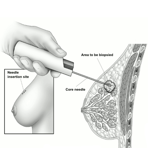
Core Needle
Biopsy
A larger hollow needle is used to obtain small tissue samples from the abnormal area.
Benefits:
- Minimally invasive
- Performed under local anesthesia
- Most commonly used breast biopsy technique
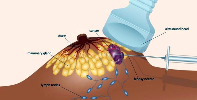
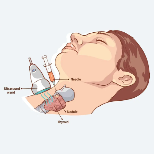
Ultrasound-Guided Breast Biopsy
Real-time ultrasound imaging guides the needle precisely to the targeted area.
Ideal For:
- Ultrasound-visible breast lesions
- Deep breast abnormalities
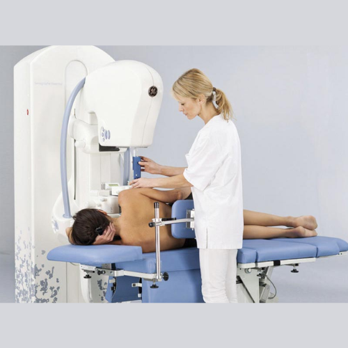
Stereotactic Breast Biopsy
Mammography imaging is used to locate and sample suspicious areas, particularly calcifications.
Ideal For:
- Microcalcifications
- Mammogram-detected abnormalities
MRI-Guided Breast Biopsy
MRI technology guides tissue sampling when abnormalities are only visible on MRI scans.
Ideal For:
- Complex breast lesions
- High-risk patients
How the Procedure Is Performed
Step 1: Evaluation
The specialist reviews your imaging studies and determines the most appropriate biopsy technique.
Step 2: Local Anesthesia
The biopsy area is numbed to ensure comfort.
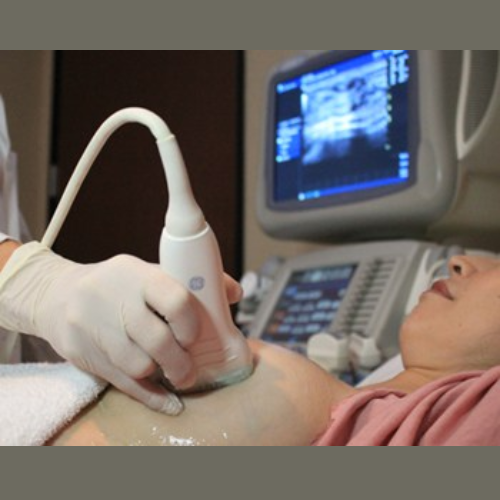
Step 3: Image Guidance
Ultrasound, mammography, or MRI is used to accurately locate the abnormal tissue.
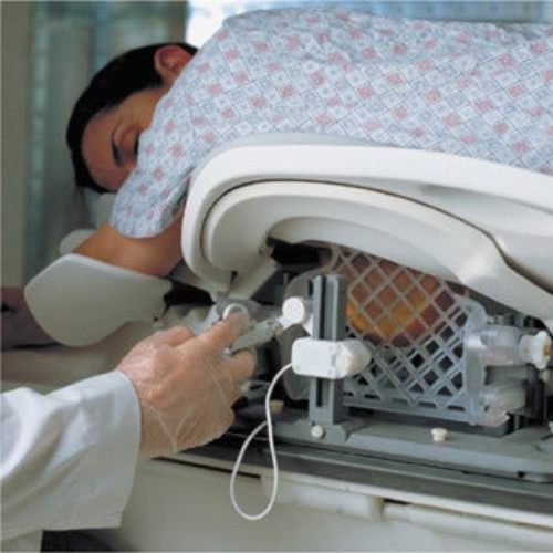
Step 4: Tissue Sampling
Small tissue samples are collected using a specialized biopsy needle.
Step 5: Laboratory Analysis
The collected tissue is sent to a pathology laboratory for detailed examination.
Benefits of Image-Guided Breast Biopsy
Minimally Invasive
No major surgery is required.
Highly Accurate
Provides reliable tissue diagnosis.
Faster Recovery
Most patients resume normal activities within 24 hours.
Minimal Scarring
Only a tiny incision or needle puncture is needed.
Outpatient Procedure
Typically completed in less than an hour.
Why Choose Our Breast Imaging & Interventional Team?
Experienced breast radiologists and interventional experts.
State-of-the-art ultrasound, mammography, and MRI-guided biopsy systems.
Accurate tissue sampling with minimal discomfort.
Compassionate support throughout the diagnostic process.
Integrated diagnosis, consultation, and treatment planning under one roof.
Frequently Asked Questions
Most breast biopsies are performed under local anesthesia, making the procedure comfortable with minimal discomfort.
The procedure usually takes 20–60 minutes depending on the biopsy type.
Most image-guided biopsies leave little to no visible scarring.
Results are typically available within 3–7 days.
No. Breast biopsy procedures are safe and do not cause cancer to spread.